What are intraoral scanners?
Digital intraoral scanners have become an ongoing trend in the dental industry and the popularity is only getting bigger. But what exactly is an intraoral scanner? Here we take a closer look at this incredible tool that makes all the difference, elevating the scanning experience for both doctors and patients to a whole new level.
What are intraoral scanners?
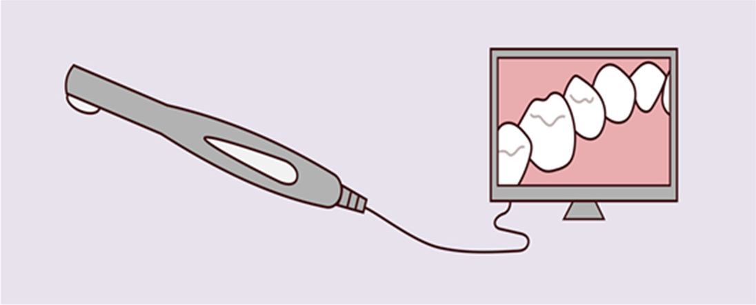
An intraoral scanner is a handheld device used to directly create digital impression data of the oral cavity. Light source from the scanner is projected onto the scan objects, such as full dental arches, and then a 3D model processed by the scanning software will be displayed in real-time on a touch screen. The device provides accurate details of the hard and soft tissues located in the oral area through high-quality images. It is becoming a more popular choice for clinics and dentists due to short lab turnaround times and excellent 3D image outputs.
What is an Intraoral Scanner and How Does it Work1
Development of Intraoral scanners
In the 18th century, methods of taking impressions and making models were already available. At that time dentists developed many impression materials such as impregum, condensation /addition silicone, agar, alginate, etc. But impression making seems error-prone and is still uncomfortable to patients and time-consuming to dentists. To overcome these limitations, intraoral digital scanners have developed as an alternative to traditional impressions.
The advent of intraoral scanners has coincided with CAD/CAM technology development, bringing many benefits to the practitioners. In the 1970s, the concept of computer-aided design/ computer-aided manufacturing (CAD/CAM) was first introduced in dental applications by Dr. Francois Duret. By 1985, the first intraoral scanner became commercially available, used by labs to fabricate precise restorations. With the introduction of the first digital scanner, dentistry was offered an exciting alternative to conventional impressions. Although the scanners of the 80s are far from the modern versions we use today, digital technology has continued to evolve over the past decade, producing scanners that are faster, more accurate and smaller than ever before.
Today, intraoral scanners and CAD/CAM technology offer easier treatment planning, more intuitive workflow, simplified learning curves, improved case acceptance, produce more accurate results, and expand the types of treatments available. No wonder more and more dental practices are realizing the need to enter the digital world— the future of dentistry.
How do intraoral scanners work?
An intraoral scanner consists of a handheld camera wand, a computer, and software. The small, smooth wand is connected to a computer that runs custom software that processes the digital data sensed by the camera. The smaller the scanning wand, the more flexible it is in reaching deep into the oral area to capture accurate and precise data. The procedure is less likely to induce gag response, making the scanning experience more comfortable for patients.
In the beginning, dentists will insert the scanning wand into the patient's mouth and gently move it over the surface area of the teeth. The wand automatically captures the size and shape of each tooth. It only takes a minute or two to scan, and the system will be able to produce a detailed digital impression. (For example, Launca DL206 intraoral scanner takes less than 40 seconds to complete a full arch scan). The dentist can view the real-time images on the computer, which can be magnified and manipulated to enhance details. The data will be transmitted to labs to fabricate any needed appliances. With this instant feedback, the whole process will be more efficient, saving time and allowing dentists to diagnose more patients.


